Virus-Like Particles (VLPs) Platform
Virus-like particles (VLPs) are hollow protein particles that are similar in shape to natural virus particles and are self-assembled by one or more structural proteins of viruses. The diameter of VLPs is about 20~200nm, which has a regular spatial structure and superior biocompatibility. The conformation of VLPs is very similar to that of real viruses, but it does not contain the nucleic acid required for virus replication and has no infective ability. Therefore, it does not have potential risks such as incomplete inactivation of virus, virus genome exchange and recombination, and virus atavism. Currently, VLPs have been widely applied for a variety of applications such as vaccines, antibody development, delivery systems, bioimaging, and cell targeting. Of note, VLPs have proven excellent utility in membrane protein display.
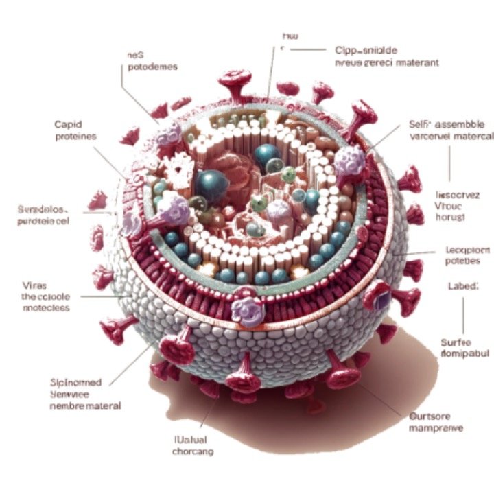
Figure 1. The diagram of a virus-like particle (VLP) structure.
Beta Lifescience has developed Virus-Like Particles (VLPs) Technology based on our magic Membrane Protein Production platform to efficiently express and purify membrane proteins.
Membrane Protein VLPs (MP-VLPs) are non-infectious virus-like particles (VLPs, about 150 nm in diameter) that display highly-expressed copies of specific membrane proteins in their native conformation. MP-VLPs are produced from host systems by co-expressing the retroviral structural core polyprotein (gag) and the target membrane protein. Retroviral gag core proteins self-assemble at the plasma membrane, where they bud from the host cells over-expressing a membrane protein of interest, enabling the formation of MP-VLPs.
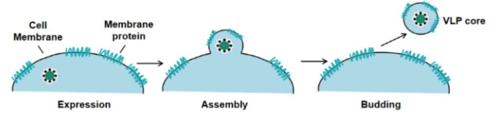
Figure 2. Schematic diagram of a VLP-expressed protein
Production Methods for the VLP Technology Platform
The appropriate choice of expression systems is one of the determinant factors in Membrane Protein Expression in VLPs. Beta Lifescience can perform VLPs Production by five following expression systems:
• Virus-like Particles (VLPs) Production in Bacterial Cells System
The bacterial cell system is the most widely used expression system for VLP production. This system is based on our well-characterized commercial Escherichia coli (E. coli) strains. The bacterial cell system not only enables VLPs to be produced in high yield and at low cost, but can also be easily scaled up. The main drawback of the bacterial cell system is the inability to perform post-translational modifications on recombinant proteins.
• Virus-like Particles (VLPs) Production in Mammalian Cells System
VLPs have shown good potential for the development of human vaccines and gene therapies. The mammalian cell system offers several advantages for VLP production. Compared with other expression systems, the mammalian cell system can not only improve production flexibility and stability, but also restore specific native glycosylation. Beta Lifescience can enable customized VLP production (particularly for influenza virus) using a wide range of mammalian cells, including HEK293 and CHO cells.
• Virus-like Particles (VLPs) Production in Yeast Cells System
The production of VLPs in yeast cells enables high-yield VLPs to be obtained in high-speed manufacturing. Compared with E. coli expression systems, yeast cells such as the Hansenula and Pichia strains are more complex, as they can select stable recombinants that contain the transgene integrated into the genome.
• Virus-like Particles (VLPs) Production in Insect Cells System
The insect cell system is another widely used system for VLP production, and offers the following advantages: rapid growth rate, easy scale-up capability and capacity for post-translational modification. The insect cell system can produce very complex VLPs, but the production yield is not very high.
• Virus-like Particles (VLPs) Production in Plant Cells System
The plant cell system is another attractive system, particularly for the production of VLP vaccines. VLPs in plant cells keep the manufacturing process low-cost, ensure appropriate post-translational modification and assembly, and reduce the risk of introducing adventitious human pathogens.
Advantages of Membrane Protein Expression in VLPs
·Ensure the natural conformation of transmembrane protein for many times, and improve the success rate of isolating antibodies that can recognize the structure of natural membrane protein
·The abundance of target antigen in encapsulated VLPs is higher than that in over expressed cells
·Higher immunogenicity
·Can be used as the best targets for dendritic cells and phage display in vivo because of their 100-300nm in size
·Suitable for immunization/ELISA/SPR/BLI/cell experiment/CAR detection
Service Details for VLP-based Membrane Protein Expression
Applications of Membrane Protein Expression in VLPs
VLPs present highly expressed copies of membrane proteins in their native conformation, providing an alternative to stable cell lines, crude membranes, detergent-solubilized proteins and other membrane protein preparation strategies (liposomes, nanodiscs). VLPs can be used for a wide range of applications.
·Immunization for antibody production
·Hybridoma screening for antibody discovery
·Phage and yeast display for antibody discovery
·Antibody characterization
·Membrane protein binding assays
·Membrane protein functional assays