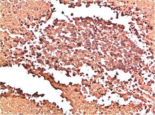
Mouse anti-Human/Mouse/Rat COL2A1 Monoclonal Antibody

Mouse anti-Human/Mouse/Rat COL2A1 Monoclonal Antibody — Type II collagen is specific for cartilaginous tissues.

SKU: BCREC-000747MA Category:

Product Specifications

Uniprot No.P02458
Target NamesCOL2A1
Species ReactivityMouse
ImmunogenSynthetic Peptide
Immunogen SpeciesHomo sapiens (Human)
ConjugateNon-conjugated
ClonalityMonoclonal
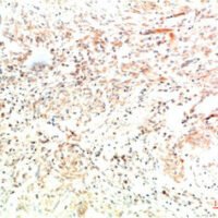
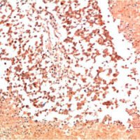
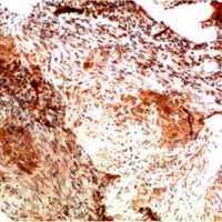
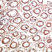
ApplicationsELISA,IHC
BufferPBS, 50% glycerol, 0.05% Proclin 300, 0.05%BSA
StorageUpon receipt, store at -20°C or -80°C. Avoid repeated freeze.

Function

Type II collagen is specific for cartilaginous tissues. It is essential for the normal embryonic development of the skeleton, for linear growth and for the ability of cartilage to resist compressive forces.

Biological Context

Subcellular Location: Secreted, extracellular space, extracellular matrix
Tissue Specificity: Isoform 2 is highly expressed in juvenile chondrocyte and low in fetal chondrocyte
Disease Association: Spondyloepiphyseal dysplasia congenital type (SEDC) : Disorder characterized by disproportionate short stature and pleiotropic involvement of the skeletal and ocular systems. [The disease is caused by variants affecting the gene represented in this entry] | Spondyloepiphyseal dysplasia, Stanescu type (SEDSTN) : An autosomal dominant spondyloepiphyseal dysplasia characterized by glycoproteins accumulation in chondrocytes. Clinical features include progressive joint contractures, premature degenerative joint disease particularly in the knee, hip and finger joints, and osseous distention of the metaphyseal ends of the phalanges causing swolling of interphalangeal joints of the hands. Radiological features include generalized platyspondyly, hypoplastic pelvis, epiphyseal flattening with metaphyseal splaying of the long bones, and enlarged phalangeal epimetaphyses of the hands. [The disease is caused by variants affecting the gene represented in this entry] | Spondyloepimetaphyseal dysplasia, Strudwick type (SEMDSTWK) : A bone disease characterized by disproportionate short stature from birth, with a very short trunk and shortened limbs, and skeletal abnormalities including lordosis, scoliosis, flattened vertebrae, pectus carinatum, coxa vara, clubfoot, and abnormal epiphyses or metaphyses. A distinctive radiographic feature is irregular sclerotic changes, described as dappled in the metaphyses of the long bones. [The disease is caused by variants affecting the gene represented in this entry] | Achondrogenesis 2 (ACG2) : An autosomal dominant disease characterized by the absence of ossification in the vertebral column, sacrum and pubic bones. [The disease is caused by variants affecting the gene represented in this entry] | Legg-Calve-Perthes disease (LCPD) : Characterized by loss of circulation to the femoral head, resulting in avascular necrosis in a growing child. Clinical pictures of the disease vary, depending on the phase of disease progression through ischemia, revascularization, fracture and collapse, and repair and remodeling of the bone. [The disease is caused by variants affecting the gene represented in this entry] | Kniest dysplasia (KD) : Moderately severe chondrodysplasia phenotype that results from mutations in the COL2A1 gene. Characteristics of the disorder include a short trunk and extremities, mid-face hypoplasia, cleft palate, myopia, retinal detachment, and hearing loss. [The disease is caused by variants affecting the gene represented in this entry] | Avascular necrosis of femoral head, primary, 1 (ANFH1) : A disease characterized by mechanical failure of the subchondral bone, and degeneration of the hip joint. It usually leads to destruction of the hip joint in the third to fifth decade of life. The clinical manifestations, such as pain on exertion, a limping gait, and a discrepancy in leg length, cause considerable disability. ANFH1 inheritance is autosomal dominant. [The disease is caused by variants affecting the gene represented in this entry] | Osteoarthritis with mild chondrodysplasia (OSCDP) : Osteoarthritis is a common disease that produces joint pain and stiffness together with radiologic evidence of progressive degeneration of joint cartilage. [The disease is caused by variants affecting the gene represented in this entry] | Platyspondylic lethal skeletal dysplasia Torrance type (PLSD-T) : Platyspondylic lethal skeletal dysplasias (PLSDs) are a heterogeneous group of chondrodysplasias characterized by severe platyspondyly and limb shortening. PLSD-T is characterized by varying platyspondyly, short ribs with anterior cupping, hypoplasia of the lower ilia with broad ischial and pubic bones, and shortening of the tubular bones with splayed and cupped metaphyses. Histology of the growth plate typically shows focal hypercellularity with slightly enlarged chondrocytes in the resting cartilage and relatively well-preserved columnar formation and ossification at the chondro-osseous junction. PLSD-T is generally a perinatally lethal disease, but a few long-term survivors have been reported. [The disease is caused by variants affecting the gene represented in this entry] | Multiple epiphyseal dysplasia with myopia and conductive deafness (EDMMD) : A generalized skeletal dysplasia associated with significant morbidity. Joint pain, joint deformity, waddling gait, and short stature are the main clinical signs and symptoms. EDMMD is an autosomal dominant disorder characterized by epiphyseal dysplasia associated with progressive myopia, retinal thinning, crenated cataracts, conductive deafness. [The disease is caused by variants affecting the gene represented in this entry] | Spondyloperipheral dysplasia (SPD) : SPD patients manifest short stature, midface hypoplasia, sensorineural hearing loss, spondyloepiphyseal dysplasia, platyspondyly and brachydactyly. [The disease is caused by variants affecting the gene represented in this entry] | Stickler syndrome 1 (STL1) : An autosomal dominant form of Stickler syndrome, an inherited disorder that associates ocular signs with more or less complete forms of Pierre Robin sequence, bone disorders and sensorineural deafness. Ocular disorders may include juvenile cataract, myopia, strabismus, vitreoretinal or chorioretinal degeneration, retinal detachment, and chronic uveitis. Pierre Robin sequence includes an opening in the roof of the mouth (a cleft palate), a large tongue (macroglossia), and a small lower jaw (micrognathia). Bones are affected by slight platyspondylisis and large, often defective epiphyses. Juvenile joint laxity is followed by early signs of arthrosis. The degree of hearing loss varies among affected individuals and may become more severe over time. Syndrome expressivity is variable. [The disease is caused by variants affecting the gene represented in this entry] | Stickler syndrome 1 non-syndromic ocular (STL1O) : An autosomal dominant form of Stickler syndrome characterized by the ocular signs typically seen in Stickler syndrome type 1 such as cataract, myopia, retinal detachment. Systemic features of premature osteoarthritis, cleft palate, hearing impairment, and craniofacial abnormalities are either absent or very mild. [The disease is caused by variants affecting the gene represented in this entry] | Rhegmatogenous retinal detachment autosomal dominant (DRRD) : A eye disease that most frequently results from a break or tear in the retina that allows fluid from the vitreous humor to enter the potential space beneath the retina. It is often associated with pathologic myopia and in most cases leads to visual impairment or blindness if untreated. [The disease is caused by variants affecting the gene represented in this entry] | Czech dysplasia (CZECHD) : A skeletal dysplasia characterized by early-onset, progressive pseudorheumatoid arthritis, platyspondyly, and short third and fourth toes. [The disease is caused by variants affecting the gene represented in this entry] | Vitreoretinopathy with phalangeal epiphyseal dysplasia (VPED) : An autosomal dominant disorder characterized by rhegmatogenous retinal detachment, premature arthropathy, and development of phalangeal epiphyseal dysplasia resulting in brachydactyly. [The disease is caused by variants affecting the gene represented in this entry] | Spondylometaphyseal dysplasia, Algerian type (SMDALG) : A form of spondylometaphyseal dysplasia, a group of short stature disorders distinguished by abnormalities in the vertebrae and the metaphyses of the tubular bones. SMDALG is an autosomal dominant form characterized by short trunk and severe genu valgum. Myopia may be a syndromic component. SMDALG radiological hallmarks include moderate platyspondyly, particularly with dorsal vertebral flattening, short ilia with narrow greater sciatic notches and generalized metaphyseal dysplasia of the long bones. The metaphyseal changes are most conspicuous in the hip and knee, and are associated with coxa vara and severe genu valgum. The short tubular bones are mildly affected. The epiphyses of the tubular bones are said to be normal. [The disease may be caused by variants affecting the gene represented in this entry]

Product Specifications

Mouse anti-Human/Mouse/Rat COL2A1 Monoclonal Antibody is a recombinant protein. Suitable for ELISA and Western Blot applications.

Catalog number: BCREC-000747MA.

SDS-PAGE: Single band at expected molecular weight confirming purity.

ELISA: Suitable as coating antigen or detection standard.

Western Blot: Compatible with standard Western Blot protocols.

Protein Interaction: Validated for SPR (Surface Plasmon Resonance) and BLI (Bio-Layer Interferometry) studies.

Shipping: Shipped at ambient temperature. Lyophilized protein is stable during transit.

Storage: Store lyophilized protein at -20°C to -80°C. Reconstituted protein should be aliquoted and stored at -80°C. Avoid repeated freeze-thaw cycles.

Shelf Life: 12 months from date of receipt when stored as recommended.

Shipping Time: Orders placed before 2 PM EST ship same day. International orders typically deliver within 5-10 business days.

Protein Biology

Function

Type II collagen is specific for cartilaginous tissues. It is essential for the normal embryonic development of the skeleton, for linear growth and for the ability of cartilage to resist compressive forces

Subcellular Location

Secreted, extracellular space, extracellular matrix

Disease Association

Spondyloepiphyseal dysplasia congenital type (SEDC) : Disorder characterized by disproportionate short stature and pleiotropic involvement of the skeletal and ocular systems. [The disease is caused by variants affecting the gene represented in this entry] | Spondyloepiphyseal dysplasia, Stanescu type (SEDSTN) : An autosomal dominant spondyloepiphyseal dysplasia characterized by glycoproteins accumulation in chondrocytes. Clinical features include progressive joint contractures, premature degenerative joint disease particularly in the knee, hip and finger joints, and osseous distention of the metaphyseal ends of the phalanges causing swolling of interphalangeal joints of the hands. Radiological features include generalized platyspondyly, hypoplastic pelvis, epiphyseal flattening with metaphyseal splaying of the long bones, and enlarged phalangeal epimetaphyses of the hands. [The disease is caused by variants affecting the gene represented in this entry] | Spondyloepimetaphyseal dysplasia, Strudwick type (SEMDSTWK) : A bone disease characterized by disproportionate short stature from birth, with a very short trunk and shortened limbs, and skeletal abnormalities including lordosis, scoliosis, flattened vertebrae, pectus carinatum, coxa vara, clubfoot, and abnormal epiphyses or metaphyses. A distinctive radiographic feature is irregular sclerotic changes, described as dappled in the metaphyses of the long bones. [The disease is caused by variants affecting the gene represented in this entry] | Achondrogenesis 2 (ACG2) : An autosomal dominant disease characterized by the absence of ossification in the vertebral column, sacrum and pubic bones. [The disease is caused by variants affecting the gene represented in this entry] | Legg-Calve-Perthes disease (LCPD) : Characterized by loss of circulation to the femoral head, resulting in avascular necrosis in a growing child. Clinical pictures of the disease vary, depending on the phase of disease progression through ischemia, revascularization, fracture and collapse, and repair and remodeling of the bone. [The disease is caused by variants affecting the gene represented in this entry] | Kniest dysplasia (KD) : Moderately severe chondrodysplasia phenotype that results from mutations in the COL2A1 gene. Characteristics of the disorder include a short trunk and extremities, mid-face hypoplasia, cleft palate, myopia, retinal detachment, and hearing loss. [The disease is caused by variants affecting the gene represented in this entry] | Avascular necrosis of femoral head, primary, 1 (ANFH1) : A disease characterized by mechanical failure of the subchondral bone, and degeneration of the hip joint. It usually leads to destruction of the hip joint in the third to fifth decade of life. The clinical manifestations, such as pain on exertion, a limping gait, and a discrepancy in leg length, cause considerable disability. ANFH1 inheritance is autosomal dominant. [The disease is caused by variants affecting the gene represented in this entry] | Osteoarthritis with mild chondrodysplasia (OSCDP) : Osteoarthritis is a common disease that produces joint pain and stiffness together with radiologic evidence of progressive degeneration of joint cartilage. [The disease is caused by variants affecting the gene represented in this entry] | Platyspondylic lethal skeletal dysplasia Torrance type (PLSD-T) : Platyspondylic lethal skeletal dysplasias (PLSDs) are a heterogeneous group of chondrodysplasias characterized by severe platyspondyly and limb shortening. PLSD-T is characterized by varying platyspondyly, short ribs with anterior cupping, hypoplasia of the lower ilia with broad ischial and pubic bones, and shortening of the tubular bones with splayed and cupped metaphyses. Histology of the growth plate typically shows focal hypercellularity with slightly enlarged chondrocytes in the resting cartilage and relatively well-preserved columnar formation and ossification at the chondro-osseous junction. PLSD-T is generally a perinatally lethal disease, but a few long-term survivors have been reported. [The disease is caused by variants affecting the gene represented in this entry] | Multiple epiphyseal dysplasia with myopia and conductive deafness (EDMMD) : A generalized skeletal dysplasia associated with significant morbidity. Joint pain, joint deformity, waddling gait, and short stature are the main clinical signs and symptoms. EDMMD is an autosomal dominant disorder characterized by epiphyseal dysplasia associated with progressive myopia, retinal thinning, crenated cataracts, conductive deafness. [The disease is caused by variants affecting the gene represented in this entry] | Spondyloperipheral dysplasia (SPD) : SPD patients manifest short stature, midface hypoplasia, sensorineural hearing loss, spondyloepiphyseal dysplasia, platyspondyly and brachydactyly. [The disease is caused by variants affecting the gene represented in this entry] | Stickler syndrome 1 (STL1) : An autosomal dominant form of Stickler syndrome, an inherited disorder that associates ocular signs with more or less complete forms of Pierre Robin sequence, bone disorders and sensorineural deafness. Ocular disorders may include juvenile cataract, myopia, strabismus, vitreoretinal or chorioretinal degeneration, retinal detachment, and chronic uveitis. Pierre Robin sequence includes an opening in the roof of the mouth (a cleft palate), a large tongue (macroglossia), and a small lower jaw (micrognathia). Bones are affected by slight platyspondylisis and large, often defective epiphyses. Juvenile joint laxity is followed by early signs of arthrosis. The degree of hearing loss varies among affected individuals and may become more severe over time. Syndrome expressivity is variable. [The disease is caused by variants affecting the gene represented in this entry] | Stickler syndrome 1 non-syndromic ocular (STL1O) : An autosomal dominant form of Stickler syndrome characterized by the ocular signs typically seen in Stickler syndrome type 1 such as cataract, myopia, retinal detachment. Systemic features of premature osteoarthritis, cleft palate, hearing impairment, and craniofacial abnormalities are either absent or very mild. [The disease is caused by variants affecting the gene represented in this entry] | Rhegmatogenous retinal detachment autosomal dominant (DRRD) : A eye disease that most frequently results from a break or tear in the retina that allows fluid from the vitreous humor to enter the potential space beneath the retina. It is often associated with pathologic myopia and in most cases leads to visual impairment or blindness if untreated. [The disease is caused by variants affecting the gene represented in this entry] | Czech dysplasia (CZECHD) : A skeletal dysplasia characterized by early-onset, progressive pseudorheumatoid arthritis, platyspondyly, and short third and fourth toes. [The disease is caused by variants affecting the gene represented in this entry] | Vitreoretinopathy with phalangeal epiphyseal dysplasia (VPED) : An autosomal dominant disorder characterized by rhegmatogenous retinal detachment, premature arthropathy, and development of phalangeal epiphyseal dysplasia resulting in brachydactyly. [The disease is caused by variants affecting the gene represented in this entry] | Spondylometaphyseal dysplasia, Algerian type (SMDALG) : A form of spondylometaphyseal dysplasia, a group of short stature disorders distinguished by abnormalities in the vertebrae and the metaphyses of the tubular bones. SMDALG is an autosomal dominant form characterized by short trunk and severe genu valgum. Myopia may be a syndromic component. SMDALG radiological hallmarks include moderate platyspondyly, particularly with dorsal vertebral flattening, short ilia with narrow greater sciatic notches and generalized metaphyseal dysplasia of the long bones. The metaphyseal changes are most conspicuous in the hip and knee, and are associated with coxa vara and severe genu valgum. The short tubular bones are mildly affected. The epiphyses of the tubular bones are said to be normal. [The disease may be caused by variants affecting the gene represented in this entry]

Tissue Specificity

Isoform 2 is highly expressed in juvenile chondrocyte and low in fetal chondrocyte

Subunit

Homotrimers of alpha 1(II) chains

Gene: COL2A1  |  Organism: Homo sapiens  |  Synonyms: Alpha-1 type II collagen
Key Publications

Frequently Asked Questions

How do I order or inquire about this product?

Fill out the Online Inquiry form with your required quantity and specifications. You can also email sales@biocrestsci.com. Our team typically responds within 4 business hours with a quote and availability confirmation.

What is the shipping and delivery time?

Orders placed before 2 PM EST ship the same day. Domestic (US) delivery typically takes 2-3 business days. International orders deliver within 5-10 business days. All products are shipped at ambient temperature with appropriate packaging to ensure stability.

How should I store this recombinant protein?

Lyophilized proteins should be stored at -20°C to -80°C upon receipt. After reconstitution, aliquot and store at -80°C. Avoid repeated freeze-thaw cycles. Shelf life is 12 months from date of receipt when stored as recommended.

What quality controls are performed on your products?

Each product undergoes SDS-PAGE purity analysis (typically >85-95%), endotoxin testing, and bioactivity validation. Products are validated for ELISA, Western Blot, and SPR/BLI applications as specified on this product page. A Certificate of Analysis (CoA) is available upon request.

Shopping Cart
Scroll to Top