, ,

Recombinant Hepatitis A Virus Cellular Receptor 2 (HAVCR2)

Recombinant Hepatitis A Virus Cellular Receptor 2 (HAVCR2) — Cell surface receptor implicated in modulating innate and adaptive immune responses. Purity >90%.

SKU: BCRETP-000045 Categories: , ,

Product Specifications

Product SkuBCRETP-000045
Product DescriptionRecombinant Hepatitis A Virus Cellular Receptor 2 (HAVCR2) is expressed from HEK293 with Fc tag at the C-Terminus. It contains 22-200aa. [Accession | Q8TDQ0].
Uniprot No.Q8TDQ0
SynonymsKIM-3; TIM3; TIMD3; Tim-3; T-cell membrane protein 3; T-cell immunoglobulin mucin receptor 3; T-cell immunoglobulin and mucin domain-containing protein 3
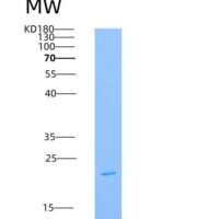
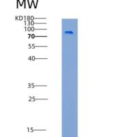
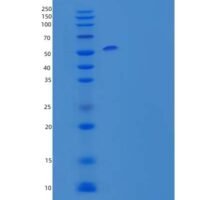
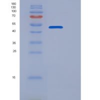
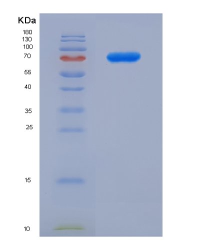
PurityGreater than 90% as determined by SDS-PAGE.
Expression SystemHEK293 Cells
Expression Region22-200aa
Tag InfoFc, 6×His tag at the C-terminus
Molecular weight80 kDa
EndotoxinLess than 1.0 EU/μg as determined by LAL method.

Function

Cell surface receptor implicated in modulating innate and adaptive immune responses. Generally accepted to have an inhibiting function. Reports on stimulating functions suggest that the activity may be influenced by the cellular context and/or the respective ligand.

Biological Context

Subcellular Location: Cell membrane (Single-pass type I membrane protein); Cell junction; Cell membrane
Tissue Specificity: Expressed in T-helper type 1 (Th1) lymphocytes. Expressed on regulatory T (Treg) cells after TCR stimulation. Expressed in dendritic cells and natural killer (NK) cells. Expressed in epithelial tissues. Expression is increased on CD4+ and CD8+ T-cells in chronic hepatitis C virus (HCV) infection. In progressive HIV-1 infection, expression is up-regulated on HIV-1-specific CD8 T-cells
Disease Association: [May be involved in T-cell exhaustion associated with chronic viral infections such as with human immunodeficiency virus (HIV) and hepatitic C virus (HCV)] | T-cell lymphoma, subcutaneous panniculitis-like (SPTCL) : An uncommon form of T-cell non-Hodgkin lymphoma, in which cytotoxic CD8+ T-cells infiltrate subcutaneous adipose tissue, and rimming adipocytes in a lace-like pattern. Affected individuals typically present with multiple subcutaneous nodules, systemic B-cell symptoms, and, in a subset of cases, autoimmune disorders, most commonly systemic lupus erythematosus. A subset of patients develop hemophagocytic lymphohistiocytosis. SPTCL transmission pattern is consistent with autosomal recessive inheritance with incomplete penetrance. [Disease susceptibility is associated with variants affecting the gene represented in this entry]

Product Specifications

Recombinant Hepatitis A Virus Cellular Receptor 2 (HAVCR2) is a recombinant protein, expressed in HEK293 Cells, covering amino acids 22-200aa, with Fc, 6×His tag at the C-terminus tag, molecular weight 80 kDa, purity Greater than 90% as determined by SDS-PAGE.. Suitable for ELISA and Western Blot applications.

SDS-PAGE: Single band at expected molecular weight confirming purity.

ELISA: Suitable as coating antigen or detection standard.

Western Blot: Compatible with standard Western Blot protocols.

Protein Interaction: Validated for SPR (Surface Plasmon Resonance) and BLI (Bio-Layer Interferometry) studies.

Shipping: Shipped at ambient temperature. Lyophilized protein is stable during transit.

Storage: Store lyophilized protein at -20°C to -80°C. Reconstituted protein should be aliquoted and stored at -80°C. Avoid repeated freeze-thaw cycles.

Shelf Life: 12 months from date of receipt when stored as recommended.

Shipping Time: Orders placed before 2 PM EST ship same day. International orders typically deliver within 5-10 business days.

Protein Biology

Function

Cell surface receptor implicated in modulating innate and adaptive immune responses. Generally accepted to have an inhibiting function. Reports on stimulating functions suggest that the activity may be influenced by the cellular context and/or the respective ligand (PubMed:24825777). Regulates macrophage activation (PubMed:11823861). Inhibits T-helper type 1 lymphocyte (Th1)-mediated auto- and alloimmune responses and promotes immunological tolerance (PubMed:14556005). In CD8+ cells attenuates TCR-induced signaling, specifically by blocking NF-kappaB and NFAT promoter activities resulting in the loss of IL-2 secretion. The function may implicate its association with LCK proposed to impair phosphorylation of TCR subunits, and/or LGALS9-dependent recruitment of PTPRC to the immunological synapse (PubMed:24337741, PubMed:26492563). In contrast, shown to activate TCR-induced signaling in T-cells probably implicating ZAP70, LCP2, LCK and FYN (By similarity). Expressed on Treg cells can inhibit Th17 cell responses (PubMed:24838857). Receptor for LGALS9 (PubMed:16286920, PubMed:24337741). Binding to LGALS9 is believed to result in suppression of T-cell responses; the resulting apoptosis of antigen-specific cells may implicate HAVCR2 phosphorylation and disruption of its association with BAG6. Binding to LGALS9 is proposed to be involved in innate immune response to intracellular pathogens. Expressed on Th1 cells interacts with LGALS9 expressed on Mycobacterium tuberculosis-infected macrophages to stimulate antibactericidal activity including IL-1 beta secretion and to restrict intracellular bacterial growth (By similarity). However, the function as receptor for LGALS9 has been challenged (PubMed:23555261). Also reported to enhance CD8+ T-cell responses to an acute infection such as by Listeria monocytogenes (By similarity). Receptor for phosphatidylserine (PtSer); PtSer-binding is calcium-dependent. May recognize PtSer on apoptotic cells leading to their phagocytosis. Mediates the engulfment of apoptotic cells by dendritic cells. Expressed on T-cells, promotes conjugation but not engulfment of apoptotic cells. Expressed on dendritic cells (DCs) positively regulates innate immune response and in synergy with Toll-like receptors promotes secretion of TNF. In tumor-infiltrating DCs suppresses nucleic acid-mediated innate immune repsonse by interaction with HMGB1 and interfering with nucleic acid-sensing and trafficking of nucleid acids to endosomes (By similarity). Expressed on natural killer (NK) cells and acts as a coreceptor to enhance IFN-gamma production in response to LGALS9 (PubMed:22323453). In contrast, shown to suppress NK cell-mediated cytotoxicity (PubMed:22383801). Negatively regulates NK cell function in LPS-induced endotoxic shock (By similarity)

Subcellular Location

Cell membrane (Single-pass type I membrane protein); Cell junction; Cell membrane

Disease Association

[May be involved in T-cell exhaustion associated with chronic viral infections such as with human immunodeficiency virus (HIV) and hepatitic C virus (HCV)] | T-cell lymphoma, subcutaneous panniculitis-like (SPTCL) : An uncommon form of T-cell non-Hodgkin lymphoma, in which cytotoxic CD8+ T-cells infiltrate subcutaneous adipose tissue, and rimming adipocytes in a lace-like pattern. Affected individuals typically present with multiple subcutaneous nodules, systemic B-cell symptoms, and, in a subset of cases, autoimmune disorders, most commonly systemic lupus erythematosus. A subset of patients develop hemophagocytic lymphohistiocytosis. SPTCL transmission pattern is consistent with autosomal recessive inheritance with incomplete penetrance. [Disease susceptibility is associated with variants affecting the gene represented in this entry]

Tissue Specificity

Expressed in T-helper type 1 (Th1) lymphocytes. Expressed on regulatory T (Treg) cells after TCR stimulation. Expressed in dendritic cells and natural killer (NK) cells. Expressed in epithelial tissues. Expression is increased on CD4+ and CD8+ T-cells in chronic hepatitis C virus (HCV) infection. In progressive HIV-1 infection, expression is up-regulated on HIV-1-specific CD8 T-cells

Subunit

Interacts with HMGB1; impairs HMGB1 binding to B-DNA and likely HMGB1-mediated innate immune response (By similarity). Interacts with BAG6 (By similarity). Interacts (phosphorylated) with PIK3R1 and PIK3R2. Interacts (not dependent on its phosphorylation status) with FYN (By similarity). Interacts (in basal state T-cells) with VAV1; AKT1/2, LCP2, ZAP70, SYK, PIK3R1, FYN, SH3BP2 and SH2D2A. Interacts (in activated T-cells) with LCK and PLCG (PubMed:24337741, PubMed:26492563). Interacts with ILF3; this interaction promotes ILF3 ubiquitination and degradation (PubMed:34110282)

Gene: HAVCR2  |  Organism: Homo sapiens  |  Synonyms: T-cell immunoglobulin and mucin domain-containing protein 3; T-cell immunoglobulin mucin receptor 3; T-cell membrane protein 3
Key Publications

Frequently Asked Questions

How do I order or inquire about this product?

Fill out the Online Inquiry form with your required quantity and specifications. You can also email sales@biocrestsci.com. Our team typically responds within 4 business hours with a quote and availability confirmation.

What is the shipping and delivery time?

Orders placed before 2 PM EST ship the same day. Domestic (US) delivery typically takes 2-3 business days. International orders deliver within 5-10 business days. All products are shipped at ambient temperature with appropriate packaging to ensure stability.

How should I store this recombinant protein?

Lyophilized proteins should be stored at -20°C to -80°C upon receipt. After reconstitution, aliquot and store at -80°C. Avoid repeated freeze-thaw cycles. Shelf life is 12 months from date of receipt when stored as recommended.

What quality controls are performed on your products?

Each product undergoes SDS-PAGE purity analysis (typically >85-95%), endotoxin testing, and bioactivity validation. Products are validated for ELISA, Western Blot, and SPR/BLI applications as specified on this product page. A Certificate of Analysis (CoA) is available upon request.

Shopping Cart
Scroll to Top