Recombinant Human Medium-wave-sensitive opsin 1 (OPN1MW) Protein

Recombinant Human Medium-wave-sensitive opsin 1 (OPN1MW) Protein — G protein-coupled photoreceptor that selectively activates G(i) proteins in response to medium-wavelength (green) light, thereby decreasing intracellular cAMP levels. Purity >85%.

SKU: BCRECP-00211TM Category:

Product Specifications

Uniprot No.P04001
Gene NamesOPN1MW
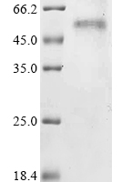
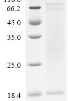
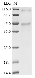
PurityGreater than 85% as determined by SDS-PAGE.
Expression Systemin vitro E.coli expression system
Expression Region1-364aa
SpeciesHomo sapiens (Human)
Tag InfoN-terminal 10xHis-tagged
Molecular weight46.6kDa
BufferIf the delivery form is liquid, the default storage buffer is Tris/PBS-based buffer, 5%-50% glycerol.If the delivery form is lyophilized powder, the buffer before lyophilization is Tris/PBS-based buffer, 6% Trehalose.
StorageStore at -20°C/-80°C upon receipt, aliquoting is necessary for mutiple use. Avoid repeated freeze-thaw cycles.

Function

G protein-coupled photoreceptor that selectively activates G(i) proteins in response to medium-wavelength (green) light, thereby decreasing intracellular cAMP levels. Activation occurs when the opsin-bound cis-retinal chromophore absorbs a photon and isomerizes to all-trans-retinal, inducing a conformational change in the opsin that triggers a G protein-mediated phototransduction cascade (Ref.6). Mediates visual perception of green light in cone photoreceptor cells.

Biological Context

Subcellular Location: Cell membrane (Multi-pass membrane protein); Photoreceptor outer segment membrane (Multi-pass membrane protein)
Tissue Specificity: The red (OPN1LW), green (OPN1MW), and blue (OPN1SW) photoreceptor pigments are found in cone cells
Disease Association: Colorblindness, partial, deutan series (CBD) : An X-linked color vision defect characterized by a dichromasy in which red and green are confused, without loss of luminance or shift or shortening of the spectrum. Dichromasy is due to the use of only two types of photoreceptors, blue plus red in deuteranopia and blue plus green in protanopia. [The disease is caused by variants affecting the gene represented in this entry] | Blue cone monochromacy (BCM) : A rare X-linked congenital stationary cone dysfunction syndrome characterized by the absence of functional long wavelength-sensitive and medium wavelength-sensitive cones in the retina. Color discrimination is severely impaired from birth, and vision is derived from the remaining preserved blue (S) cones and rod photoreceptors. BCM typically presents with reduced visual acuity, pendular nystagmus, and photophobia. Patients often have myopia. [The disease is caused by variants affecting the gene represented in this entry] | Cone dystrophy 5 (COD5) : An X-linked cone dystrophy. Cone dystrophies are retinal dystrophies characterized by progressive degeneration of the cone photoreceptors with preservation of rod function, as indicated by electroretinogram. However, some rod involvement may be present in some cone dystrophies, particularly at late stage. Affected individuals suffer from photophobia, loss of visual acuity, color vision and central visual field. Another sign is the absence of macular lesions for many years. Cone dystrophies are distinguished from the cone-rod dystrophies in which some loss of peripheral vision also occurs. [The disease is caused by variants affecting the gene represented in this entry]

Product Specifications

Recombinant Human Medium-wave-sensitive opsin 1 (OPN1MW) Protein is a recombinant protein from Homo sapiens (Human), expressed in in vitro E.coli expression system, covering amino acids 1-364aa, with N-terminal 10xHis-tagged tag, molecular weight 46.6kDa, purity Greater than 85% as determined by SDS-PAGE.. Suitable for ELISA and Western Blot applications.
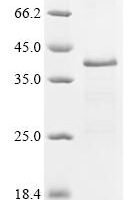
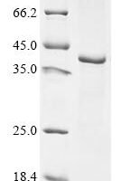
SDS-PAGE: Single band at expected molecular weight confirming purity.

ELISA: Suitable as coating antigen or detection standard.

Western Blot: Compatible with standard Western Blot protocols.

Protein Interaction: Validated for SPR (Surface Plasmon Resonance) and BLI (Bio-Layer Interferometry) studies.

Shipping: Shipped at ambient temperature. Lyophilized protein is stable during transit.

Storage: Store lyophilized protein at -20°C to -80°C. Reconstituted protein should be aliquoted and stored at -80°C. Avoid repeated freeze-thaw cycles.

Shelf Life: 12 months from date of receipt when stored as recommended.

Shipping Time: Orders placed before 2 PM EST ship same day. International orders typically deliver within 5-10 business days.

Protein Biology

Function

G protein-coupled photoreceptor that selectively activates G(i) proteins in response to medium-wavelength (green) light, thereby decreasing intracellular cAMP levels (PubMed:2937147, PubMed:6140680, Ref.6). Activation occurs when the opsin-bound cis-retinal chromophore absorbs a photon and isomerizes to all-trans-retinal, inducing a conformational change in the opsin that triggers a G protein-mediated phototransduction cascade (Ref.6). Mediates visual perception of green light in cone photoreceptor cells (PubMed:12051694, PubMed:1302020, PubMed:8666378)

Subcellular Location

Cell membrane (Multi-pass membrane protein); Photoreceptor outer segment membrane (Multi-pass membrane protein)

Disease Association

Colorblindness, partial, deutan series (CBD) : An X-linked color vision defect characterized by a dichromasy in which red and green are confused, without loss of luminance or shift or shortening of the spectrum. Dichromasy is due to the use of only two types of photoreceptors, blue plus red in deuteranopia and blue plus green in protanopia. [The disease is caused by variants affecting the gene represented in this entry] | Blue cone monochromacy (BCM) : A rare X-linked congenital stationary cone dysfunction syndrome characterized by the absence of functional long wavelength-sensitive and medium wavelength-sensitive cones in the retina. Color discrimination is severely impaired from birth, and vision is derived from the remaining preserved blue (S) cones and rod photoreceptors. BCM typically presents with reduced visual acuity, pendular nystagmus, and photophobia. Patients often have myopia. [The disease is caused by variants affecting the gene represented in this entry] | Cone dystrophy 5 (COD5) : An X-linked cone dystrophy. Cone dystrophies are retinal dystrophies characterized by progressive degeneration of the cone photoreceptors with preservation of rod function, as indicated by electroretinogram. However, some rod involvement may be present in some cone dystrophies, particularly at late stage. Affected individuals suffer from photophobia, loss of visual acuity, color vision and central visual field. Another sign is the absence of macular lesions for many years. Cone dystrophies are distinguished from the cone-rod dystrophies in which some loss of peripheral vision also occurs. [The disease is caused by variants affecting the gene represented in this entry]

Tissue Specificity

The red (OPN1LW), green (OPN1MW), and blue (OPN1SW) photoreceptor pigments are found in cone cells

Subunit

Monomer. Homodimer. Homotetramer

Gene: OPN1MW  |  Organism: Homo sapiens  |  Synonyms: Green cone photoreceptor pigment; Green-sensitive opsin
Key Publications

Frequently Asked Questions

How do I order or inquire about this product?

Fill out the Online Inquiry form with your required quantity and specifications. You can also email sales@biocrestsci.com. Our team typically responds within 4 business hours with a quote and availability confirmation.

What is the shipping and delivery time?

Orders placed before 2 PM EST ship the same day. Domestic (US) delivery typically takes 2-3 business days. International orders deliver within 5-10 business days. All products are shipped at ambient temperature with appropriate packaging to ensure stability.

How should I store this recombinant protein?

Lyophilized proteins should be stored at -20°C to -80°C upon receipt. After reconstitution, aliquot and store at -80°C. Avoid repeated freeze-thaw cycles. Shelf life is 12 months from date of receipt when stored as recommended.

What quality controls are performed on your products?

Each product undergoes SDS-PAGE purity analysis (typically >85-95%), endotoxin testing, and bioactivity validation. Products are validated for ELISA, Western Blot, and SPR/BLI applications as specified on this product page. A Certificate of Analysis (CoA) is available upon request.

Shopping Cart
Scroll to Top