Recombinant Human Melanocyte protein PMEL (PMEL), partial

Recombinant Human Melanocyte protein PMEL (PMEL), partial — Represents a potent melanoma-specific antigen. Purity >90%.

SKU: BCRECP-001407 Category:

Product Specifications

Product SkuBCRECP-001407
Product DescriptionRecombinant Human Melanocyte protein PMEL (PMEL) Protein is expressed from E.coli with N-terminal 6xHis-SUMO-tagged. It contains 25-467aa. [Accession | P40967].
Uniprot No.P40967
Gene NamesPMEL
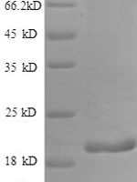
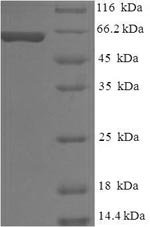
PurityGreater than 90% as determined by SDS-PAGE.
Expression SystemE.coli
Expression Region25-467aa
SpeciesHomo sapiens (Human)
Tag InfoN-terminal 6xHis-SUMO-tagged
Molecular weight63.0kDa
ActivityPlease contact us to obtain bioactivity data.
BufferIf the delivery form is liquid, the default storage buffer is Tris/PBS-based buffer, 5%-50% glycerol. If the delivery form is lyophilized powder, the buffer before lyophilization is Tris/PBS-based buffer, 6% Trehalose.
StorageStore at -20°C/-80°C upon receipt, aliquoting is necessary for mutiple use. Avoid repeated freeze-thaw cycles.
Research AreasMetabolism

Function

Represents a potent melanoma-specific antigen. Among melanoma non-mutated self-peptides, G9-154 (KTWGQYWQV), G9-209 (ITDQVPFSV) and G9-280 (YLEPGPVTA), appear to act as immunodominant common epitopes that stimulate anti-tumor immune response mediated by HLA-A-restricted cytotoxic T cells.

Biological Context

Subcellular Location: Endoplasmic reticulum membrane (Single-pass type I membrane protein); Golgi apparatus, cis-Golgi network membrane (Single-pass type I membrane protein); Endosome, multivesicular body; Melanosome; Extracellular vesicle; Secreted
Tissue Specificity: Normally expressed at low levels in quiescent adult melanocytes but overexpressed by proliferating neonatal melanocytes and during tumor growth. Overexpressed in melanomas. Some expression was found in dysplastic nevi

Product Specifications

Recombinant Human Melanocyte protein PMEL (PMEL), partial is a recombinant protein from Homo sapiens (Human), expressed in E.coli, covering amino acids 25-467aa, with N-terminal 6xHis-SUMO-tagged tag, molecular weight 63.0kDa, purity Greater than 90% as determined by SDS-PAGE.. Suitable for ELISA and Western Blot applications.
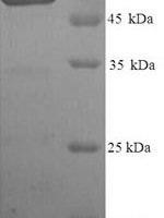
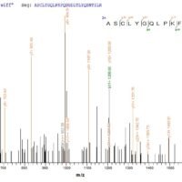
SDS-PAGE: Single band at expected molecular weight confirming purity.

ELISA: Suitable as coating antigen or detection standard.

Western Blot: Compatible with standard Western Blot protocols.

Protein Interaction: Validated for SPR (Surface Plasmon Resonance) and BLI (Bio-Layer Interferometry) studies.

Shipping: Shipped at ambient temperature. Lyophilized protein is stable during transit.

Storage: Store lyophilized protein at -20°C to -80°C. Reconstituted protein should be aliquoted and stored at -80°C. Avoid repeated freeze-thaw cycles.

Shelf Life: 12 months from date of receipt when stored as recommended.

Shipping Time: Orders placed before 2 PM EST ship same day. International orders typically deliver within 5-10 business days.

Protein Biology

Function

Represents a potent melanoma-specific antigen. Among melanoma non-mutated self-peptides, G9-154 (KTWGQYWQV), G9-209 (ITDQVPFSV) and G9-280 (YLEPGPVTA), appear to act as immunodominant common epitopes that stimulate anti-tumor immune response mediated by HLA-A-restricted cytotoxic T cells

Subcellular Location

Endoplasmic reticulum membrane (Single-pass type I membrane protein); Golgi apparatus, cis-Golgi network membrane (Single-pass type I membrane protein); Endosome, multivesicular body; Melanosome; Extracellular vesicle; Secreted

Tissue Specificity

Normally expressed at low levels in quiescent adult melanocytes but overexpressed by proliferating neonatal melanocytes and during tumor growth. Overexpressed in melanomas. Some expression was found in dysplastic nevi

Subunit

Homodimer; disulfide-linked. Dimerization in the endoplasmic reticulum and early Golgi prevents premature fibril formation. The dimers are resolved to monomers in late- or post-Golgi compartments (PubMed:26694611). Heterooligomer; amyloid-type. Processed amyloidogenic fragments assemble into fibrils that further organize into beta-sheet quaternary amyloid structures (PubMed:28272432, PubMed:30988362). Interacts (via luminal C-terminal fragment) with CD63; this is important for sorting of the luminal fragment in tetraspanin rich microdomains in stage I melanosomes to prevent premature lysosomal degradation (PubMed:21962903). Interacts with APOE; this allows the loading of the luminal fragment on ILVs to induce fibril nucleation (PubMed:26387950). Interacts with MLANA (PubMed:15695812)

Gene: PMEL  |  Organism: Homo sapiens  |  Synonyms: ME20-M; Melanocyte protein Pmel 17; Melanocytes lineage-specific antigen GP100; Melanoma-associated ME20 antigen; P1; P100; Premelanosome protein; Silver locus protein homolog
Key Publications

Frequently Asked Questions

How do I order or inquire about this product?

Fill out the Online Inquiry form with your required quantity and specifications. You can also email sales@biocrestsci.com. Our team typically responds within 4 business hours with a quote and availability confirmation.

What is the shipping and delivery time?

Orders placed before 2 PM EST ship the same day. Domestic (US) delivery typically takes 2-3 business days. International orders deliver within 5-10 business days. All products are shipped at ambient temperature with appropriate packaging to ensure stability.

How should I store this recombinant protein?

Lyophilized proteins should be stored at -20°C to -80°C upon receipt. After reconstitution, aliquot and store at -80°C. Avoid repeated freeze-thaw cycles. Shelf life is 12 months from date of receipt when stored as recommended.

What quality controls are performed on your products?

Each product undergoes SDS-PAGE purity analysis (typically >85-95%), endotoxin testing, and bioactivity validation. Products are validated for ELISA, Western Blot, and SPR/BLI applications as specified on this product page. A Certificate of Analysis (CoA) is available upon request.

Shopping Cart
Scroll to Top