Recombinant Human Midkine protein (MDK)

Recombinant Human Midkine protein (MDK) — Secreted protein that functions as a cytokine and growth factor and mediates its signal through cell-surface proteoglycan and non-proteoglycan receptors. Purity >97%.

SKU: BCACP-000886 Category:
Product SKUBCACP-000886
Product DescriptionRecombinant Human Midkine protein (MDK) protein is expressed from E.Coli with Tag-Free. It contains 21-143aa. [Accession | P21741].
Uniprot No.P21741
Gene NamesMDK
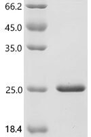
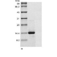
Purity>97% as determined by SDS-PAGE.
Expression SystemE.Coli
Expression Region21-143aa
SpeciesHomo sapiens (Human)
Tag InfoTag-Free
Molecular weight13.4 kDa
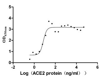
ActivityFully biologically active when compared to standard. The ED50 as determined by anti-microbial activity against E.coli. is less than 30 μg/ml, corresponding to a specific activity of >33.3 IU/mg.
BufferLyophilized from a 0.2 μm filtered 20 mM Tris-HCl, 0.5 M NaCl, 6% Trehalose, pH 8.0
StorageStore at -20°C/-80°C upon receipt, aliquoting is necessary for mutiple use. Avoid repeated freeze-thaw cycles.
Alternative NamesAmphiregulin associated protein; Amphiregulin-associated protein; ARAP; FLJ27379; Mdk; Midgestation and kidney protein; Midkine; MK 1; MK; MK_HUMAN; MK1; NEGF 2; NEGF2; Neurite growth promoting factor 2; Neurite outgrowth promoting protein; Neurite outgrowth-promoting factor 2; Neurite outgrowth-promoting protein; Retinoic acid inducible factor

Function

Secreted protein that functions as a cytokine and growth factor and mediates its signal through cell-surface proteoglycan and non-proteoglycan receptors. Binds cell-surface proteoglycan receptors via their chondroitin sulfate (CS) groups. Thereby regulates many processes like inflammatory response, cell proliferation, cell adhesion, cell growth, cell survival, tissue regeneration, cell differentiation and cell migration.

Biological Context

Subcellular Location: Secreted
Tissue Specificity: Expressed in various tumor cell lines. In insulinoma tissue predominantly expressed in precancerous lesions

Product Specifications

Recombinant Human Midkine protein (MDK) is a recombinant protein from Homo sapiens (Human), expressed in E.Coli, covering amino acids 21-143aa, with Tag-Free tag, molecular weight 13.4 kDa, purity >97% as determined by SDS-PAGE.. Suitable for ELISA and Western Blot applications.
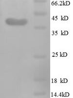
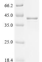
SDS-PAGE: Single band at expected molecular weight confirming purity.

ELISA: Suitable as coating antigen or detection standard.

Western Blot: Compatible with standard Western Blot protocols.

Protein Interaction: Validated for SPR (Surface Plasmon Resonance) and BLI (Bio-Layer Interferometry) studies.

Shipping: Shipped at ambient temperature. Lyophilized protein is stable during transit.

Storage: Store lyophilized protein at -20°C to -80°C. Reconstituted protein should be aliquoted and stored at -80°C. Avoid repeated freeze-thaw cycles.

Shelf Life: 12 months from date of receipt when stored as recommended.

Shipping Time: Orders placed before 2 PM EST ship same day. International orders typically deliver within 5-10 business days.

Protein Biology

Function

Secreted protein that functions as a cytokine and growth factor and mediates its signal through cell-surface proteoglycan and non-proteoglycan receptors (PubMed:10212223, PubMed:10772929, PubMed:12084985, PubMed:12122009, PubMed:12573468, PubMed:15466886, PubMed:18469519, PubMed:24458438). Binds cell-surface proteoglycan receptors via their chondroitin sulfate (CS) groups (PubMed:10212223, PubMed:12084985). Thereby regulates many processes like inflammatory response, cell proliferation, cell adhesion, cell growth, cell survival, tissue regeneration, cell differentiation and cell migration (PubMed:10212223, PubMed:10683378, PubMed:10772929, PubMed:12084985, PubMed:12122009, PubMed:12573468, PubMed:15466886, PubMed:22323540, PubMed:24458438). Participates in inflammatory processes by exerting two different activities. Firstly, mediates neutrophils and macrophages recruitment to the sites of inflammation both by direct action by cooperating namely with ITGB2 via LRP1 and by inducing chemokine expression (PubMed:10683378, PubMed:24458438). This inflammation can be accompanied by epithelial cell survival and smooth muscle cell migration after renal and vessel damage, respectively (PubMed:10683378). Secondly, suppresses the development of tolerogenic dendric cells thereby inhibiting the differentiation of regulatory T cells and also promote T cell expansion through NFAT signaling and Th1 cell differentiation (PubMed:22323540). Promotes tissue regeneration after injury or trauma. After heart damage negatively regulates the recruitment of inflammatory cells and mediates cell survival through activation of anti-apoptotic signaling pathways via MAPKs and AKT pathways through the activation of angiogenesis (By similarity). Also facilitates liver regeneration as well as bone repair by recruiting macrophage at trauma site and by promoting cartilage development by facilitating chondrocyte differentiation (By similarity). Plays a role in brain by promoting neural precursor cells survival and growth through interaction with heparan sulfate proteoglycans (By similarity). Binds PTPRZ1 and promotes neuronal migration and embryonic neurons survival (PubMed:10212223). Binds SDC3 or GPC2 and mediates neurite outgrowth and cell adhesion (PubMed:12084985, PubMed:1768439). Binds chondroitin sulfate E and heparin leading to inhibition of neuronal cell adhesion induced by binding with GPC2 (PubMed:12084985). Binds CSPG5 and promotes elongation of oligodendroglial precursor-like cells (By similarity). Also binds ITGA6:ITGB1 complex; this interaction mediates MDK-induced neurite outgrowth (PubMed:15466886, PubMed:1768439). Binds LRP1; promotes neuronal survival (PubMed:10772929). Binds ITGA4:ITGB1 complex; this interaction mediates MDK-induced osteoblast cells migration through PXN phosphorylation (PubMed:15466886). Binds anaplastic lymphoma kinase (ALK) which induces ALK activation and subsequent phosphorylation of the insulin receptor substrate (IRS1), followed by the activation of mitogen-activated protein kinase (MAPK) and PI3-kinase, and the induction of cell proliferation (PubMed:12122009). Promotes epithelial to mesenchymal transition through interaction with NOTCH2 (PubMed:18469519). During arteriogenesis, plays a role in vascular endothelial cell proliferation by inducing VEGFA expression and release which in turn induces nitric oxide synthase expression. Moreover activates vasodilation through nitric oxide synthase activation (By similarity). Negatively regulates bone formation in response to mechanical load by inhibiting Wnt/beta-catenin signaling in osteoblasts (By similarity). In addition plays a role in hippocampal development, working memory, auditory response, early fetal adrenal gland development and the female reproductive system (By similarity)

Subcellular Location

Secreted

Tissue Specificity

Expressed in various tumor cell lines. In insulinoma tissue predominantly expressed in precancerous lesions

Subunit

Homodimer. Interacts with ALK (PubMed:12122009). Interacts with LRP1; promotes neuronal survival (PubMed:10772929). Interacts with LRP2 (PubMed:10772929). Interacts with NCAM1 (PubMed:10772929). Interacts (via C-terminal) with PTPRZ1 (via chondroitin sulfate chains); this interaction is inhibited by PTN; this interaction promotes neuronal migration (PubMed:10212223). Interacts with NCL; this interaction promotes NCL clustering and lateral movements of this complex into lipid rafts leading to MDK internalization (PubMed:12147681). Interacts with LRP6 and LRP8: this interaction is calcium dependent (PubMed:12573468). Interacts with ITGA4 (PubMed:15466886). Interacts with ITGA6 (PubMed:15466886). Interacts with ITGB1 (PubMed:15466886). Interacts with ITGA4:ITGB1 complex; this interaction mediates MDK-induced osteoblast cells migration through PXN phosphorylation (PubMed:15466886). Interacts with ITGA6:ITGB1 complex; this interaction mediates MDK-induced neurite outgrowth (PubMed:15466886). Interacts with NOTCH2; this interaction mediates a nuclear accumulation of NOTCH2 and therefore activation of NOTCH2 signaling leading to interaction between HES1 and STAT3 (PubMed:18469519). Interacts with GPC2 (via heparan sulfate chain); this interaction is inhibited by heparin followed by chondroitin sulfate E; this interaction induces GPC2 clustering through heparan sulfate chain; this interaction induces neuronal cell adhesion and neurite outgrowth (PubMed:12084985). Interacts with SDC3; this interaction induces SDC3 clustering; this interaction induces neuronal cell adhesion and neurite outgrowth (PubMed:12084985). Interacts with SDC1 (By similarity). Interacts with CSPG5; this interaction promotes elongation of oligodendroglial precursor-like cells (By similarity)

Gene: MDK  |  Organism: Homo sapiens  |  Synonyms: Amphiregulin-associated protein; Midgestation and kidney protein; Neurite outgrowth-promoting factor 2; Neurite outgrowth-promoting protein
Key Publications

Frequently Asked Questions

How do I order or inquire about this product?

Fill out the Online Inquiry form with your required quantity and specifications. You can also email sales@biocrestsci.com. Our team typically responds within 4 business hours with a quote and availability confirmation.

What is the shipping and delivery time?

Orders placed before 2 PM EST ship the same day. Domestic (US) delivery typically takes 2-3 business days. International orders deliver within 5-10 business days. All products are shipped at ambient temperature with appropriate packaging to ensure stability.

How should I store this recombinant protein?

Lyophilized proteins should be stored at -20°C to -80°C upon receipt. After reconstitution, aliquot and store at -80°C. Avoid repeated freeze-thaw cycles. Shelf life is 12 months from date of receipt when stored as recommended.

What quality controls are performed on your products?

Each product undergoes SDS-PAGE purity analysis (typically >85-95%), endotoxin testing, and bioactivity validation. Products are validated for ELISA, Western Blot, and SPR/BLI applications as specified on this product page. A Certificate of Analysis (CoA) is available upon request.

Shopping Cart
Scroll to Top