Function
Acts as a guanine-nucleotide releasing factor (GEF) for RAB8A and RAB37 by promoting the conversion of inactive RAB-GDP to the active form RAB-GTP. GEF activity towards RAB8A may facilitate ciliary trafficking by modulating ciliary intracellular localization of RAB8A. GEF activity towards RAB37 maintains autophagic homeostasis and retinal function.
Biological Context
Subcellular Location: Cytoplasm, cytoskeleton, microtubule organizing center, centrosome; Cytoplasm, cytoskeleton, cilium basal body; Cytoplasm, cytoskeleton, cilium axoneme
Tissue Specificity: Heart, brain, placenta, lung, liver, muscle, kidney, retina, pancreas and fetal retinal pigment epithelium. Isoform 3 is found only in the retina. Colocalizes with RPGRIP1 in the outer segment of rod photoreceptors and cone outer segments
Disease Association: Retinitis pigmentosa 3 (RP3) : An X-linked retinal dystrophy belonging to the group of pigmentary retinopathies. Retinitis pigmentosa is characterized by retinal pigment deposits visible on fundus examination and primary loss of rod photoreceptor cells followed by secondary loss of cone photoreceptors. Patients typically have night vision blindness and loss of midperipheral visual field. As their condition progresses, they lose their far peripheral visual field and eventually central vision as well. In RP3, affected males have a severe phenotype, and carrier females show a wide spectrum of clinical features ranging from completely asymptomatic to severe retinitis pigmentosa. Heterozygous women can manifest a form of choroidoretinal degeneration which is distinguished from other types by the absence of visual defects in the presence of a brilliant, scintillating, golden-hued, patchy appearance most striking around the macula, called a tapetal-like retinal reflex. [The disease is caused by variants affecting the gene represented in this entry] | Retinitis pigmentosa, X-linked, and sinorespiratory infections with or without deafness (RPSRDF) : A disease characterized by the association of retinitis pigmentosa with recurrent upper and lower airway infections. Some patients also develop progressive hearing loss. [The disease is caused by variants affecting the gene represented in this entry] | Cone-rod dystrophy, X-linked 1 (CORDX1) : An inherited retinal dystrophy characterized by retinal pigment deposits visible on fundus examination, predominantly in the macular region, and initial loss of cone photoreceptors followed by rod degeneration. This leads to decreased visual acuity and sensitivity in the central visual field, followed by loss of peripheral vision. Severe loss of vision occurs earlier than in retinitis pigmentosa. In cone-rod dystrophy X-linked type 1 the degree of rod-photoreceptor involvement can be variable, with degeneration increasing as the disease progresses. Affected individuals (essentially all of whom are males) present with decreased visual acuity, myopia, photophobia, abnormal color vision, full peripheral visual fields, decreased photopic electroretinographic responses, and granularity of the macular retinal pigment epithelium. Although penetrance appears to be nearly 100%, there is variable expressivity with respect to age at onset and severity of symptoms. [The disease is caused by variants affecting the gene represented in this entry] | Macular degeneration, atrophic, X-linked (MDXLA) : An ocular disorder characterized by macular atrophy causing progressive loss of visual acuity with minimal peripheral visual impairment. Some patients manifest extensive macular degeneration plus peripheral loss of retinal pigment epithelium and choriocapillaries. Full-field electroretinograms (ERGs) show normal cone and rod responses in some affected males despite advanced macular degeneration. [The disease is caused by variants affecting the gene represented in this entry]
Product Specifications
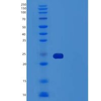
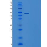
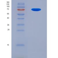
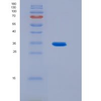
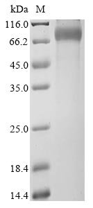
Recombinant Human X-linked retinitis pigmentosa GTPase regulator (RPGR), partial is a recombinant protein from Homo sapiens (Human), expressed in Mammalian cell, covering amino acids 54-367aa, with C-terminal hFc1-tagged tag, molecular weight 62.8kDa, purity Greater than 90% as determined by SDS-PAGE.. Suitable for ELISA and Western Blot applications.